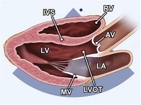
m-mode lv echocardiography plax | plax echocardiogram m-mode lv echocardiography plax Assessment of LV function with M-mode or 2-dimensional (2-D) echocardiography (Figure 2A) can be performed in the parasternal long- and short-axis views by .
profesionĀla matu kosmĒtika, frizieru instrumenti un frizĒtavu aprĪkojums . facebook @mowanmegix.lv: bondplex™ – 21. gadsimta sasniegums: nĒ! matu bojĀjumiem: facebook @reddot.lv: brauc pie mums - pĒrc lĒtĀk! -----> atlaides lĪdz 28.06.2024 (pdf .
0 · plax echocardiogram
1 · parasternal plax view echocardiogram
2 · m-mode of mv
I was planning to do a filter & fluid change and fill it with idemitsu TLS-LV (WS). Label says “customized formulation engineered for Asian vehicle models with Type-WS transmission specifications” Questions: 1. Is Idemitsu TLS-LV acceptable for the RAV? Its designation and labeling has me a little confused. 2.
PLAX view. The parasternal long axis view (PLAX) is obtained with the transducer image marker directed toward the patient’s right ear and the sound beam directed to the spine. Slight .
It is used to guide M-Mode echocardiography for left ventricular measurements. Initially the parasternal long axis view is obtained. When satisfactory images are available after .How to measure the Ejection Fraction of left ventricle using m-mode or motion mode on parasternal long axis view.THE AMERICAN SOCIETY OF ECHOCARDIOGRAPHY RECOMMENDATIONS FOR CARDIAC CHAMBER QUANTIFICATION IN ADULTS: A QUICK REFERENCE GUIDE FROM THE ASE .VI. M-Mode Measurements This section provides guidance on selected M-mode measurements. VII. Color Doppler Imaging This section defines the basic imaging windows, display, and mea .
Assessment of LV function with M-mode or 2-dimensional (2-D) echocardiography (Figure 2A) can be performed in the parasternal long- and short-axis views by .
Use the zoom function in the PLAX view for optimal visualization of LV outflow tract (LVOT) and the aortic valve with visualization of AV cusp insertion points (annulus). Both MV leaflets and .The M-mode from the LV at the mitral valve leaflet level may be useful to measurement: a) the diastolic inter-ventricular septum, b) the diastolic posterior wall of the LV. These may be .
emma chamberlain louis vuitton outfit
plax echocardiogram
PLAX M-mode: MV E-Septal separation (EPSS) EPSS is defined as the minimal distance between E point (most anterior motion of the AML during diastole) and a line tangential to the . M-mode imaging in the parasternal views will further elucidate mitral leaflet motion and define the duration of mitral-septal contact. Color Doppler and PW Doppler mapping should be integrated in the assessment of .PLAX view. The parasternal long axis view (PLAX) is obtained with the transducer image marker directed toward the patient’s right ear and the sound beam directed to the spine. Slight adjustments in angle and rotation maybe necessary to demonstrate all . It is used to guide M-Mode echocardiography for left ventricular measurements. Initially the parasternal long axis view is obtained. When satisfactory images are available after fine adjustments of the transducer position, M-Mode cursor is placed in such a .
How to measure the Ejection Fraction of left ventricle using m-mode or motion mode on parasternal long axis view.THE AMERICAN SOCIETY OF ECHOCARDIOGRAPHY RECOMMENDATIONS FOR CARDIAC CHAMBER QUANTIFICATION IN ADULTS: A QUICK REFERENCE GUIDE FROM THE ASE WORKFLOW AND LAB MANAGEMENT TASK FORCE. Accurate and reproducible assessment of cardiac chamber size and function is essential for clinical care. A standardized methodology .
VI. M-Mode Measurements This section provides guidance on selected M-mode measurements. VII. Color Doppler Imaging This section defines the basic imaging windows, display, and mea-surements for color Doppler imaging (CDI) to be integrated into the comprehensive transthoracic examination. Similarly, display of colorAssessment of LV function with M-mode or 2-dimensional (2-D) echocardiography (Figure 2A) can be performed in the parasternal long- and short-axis views by placing the calipers perpendicular to the ventricular long axis. Change in LV cavity dimensions during systole can be used to calculate LV fractional shortening and ejection fraction.
Use the zoom function in the PLAX view for optimal visualization of LV outflow tract (LVOT) and the aortic valve with visualization of AV cusp insertion points (annulus). Both MV leaflets and 2 of the 3 aortic leaflets should be visible in good quality.PLAX M-mode: MV E-Septal separation (EPSS) EPSS is defined as the minimal distance between E point (most anterior motion of the AML during diastole) and a line tangential to the most posterior excursion of the IVS within the same cardiac cycle
M-mode imaging in the parasternal views will further elucidate mitral leaflet motion and define the duration of mitral-septal contact. Color Doppler and PW Doppler mapping should be integrated in the assessment of obstruction and, when present, determine the . Rapidly moving structures such as the aortic valve and mitral valve, and endocardium have characteristic movements in M-mode. M-mode also has a great spatial resolution, which is useful for measuring ventricular dimensions in systole and diastole.PLAX view. The parasternal long axis view (PLAX) is obtained with the transducer image marker directed toward the patient’s right ear and the sound beam directed to the spine. Slight adjustments in angle and rotation maybe necessary to demonstrate all .
It is used to guide M-Mode echocardiography for left ventricular measurements. Initially the parasternal long axis view is obtained. When satisfactory images are available after fine adjustments of the transducer position, M-Mode cursor is placed in such a .How to measure the Ejection Fraction of left ventricle using m-mode or motion mode on parasternal long axis view.THE AMERICAN SOCIETY OF ECHOCARDIOGRAPHY RECOMMENDATIONS FOR CARDIAC CHAMBER QUANTIFICATION IN ADULTS: A QUICK REFERENCE GUIDE FROM THE ASE WORKFLOW AND LAB MANAGEMENT TASK FORCE. Accurate and reproducible assessment of cardiac chamber size and function is essential for clinical care. A standardized methodology .VI. M-Mode Measurements This section provides guidance on selected M-mode measurements. VII. Color Doppler Imaging This section defines the basic imaging windows, display, and mea-surements for color Doppler imaging (CDI) to be integrated into the comprehensive transthoracic examination. Similarly, display of color
Assessment of LV function with M-mode or 2-dimensional (2-D) echocardiography (Figure 2A) can be performed in the parasternal long- and short-axis views by placing the calipers perpendicular to the ventricular long axis. Change in LV cavity dimensions during systole can be used to calculate LV fractional shortening and ejection fraction.Use the zoom function in the PLAX view for optimal visualization of LV outflow tract (LVOT) and the aortic valve with visualization of AV cusp insertion points (annulus). Both MV leaflets and 2 of the 3 aortic leaflets should be visible in good quality.PLAX M-mode: MV E-Septal separation (EPSS) EPSS is defined as the minimal distance between E point (most anterior motion of the AML during diastole) and a line tangential to the most posterior excursion of the IVS within the same cardiac cycle
M-mode imaging in the parasternal views will further elucidate mitral leaflet motion and define the duration of mitral-septal contact. Color Doppler and PW Doppler mapping should be integrated in the assessment of obstruction and, when present, determine the .
costum louis vuitton

parasternal plax view echocardiogram
coque telephone louis vuitton iphone 11
m-mode of mv
Gaisma no Zemes līdz Mēnesim nonāk aptuveni 1,2 sekundēs. Gaismas ātrums vakuumā ir 299 792 458 m/s.Tā ir fizikāla konstante.Bieži dažādos aprēķinos gaismas ātrumu noapaļo uz 300 000 000 jeb 3×10 8 m/s. Optiski aktīvās vidēs gaismas ātrums samazinās, piemēram, ūdenī gaismas ātrums ir aptuveni 2,26×10 8 m/s. Izmantojot gaismas ātrumu, .Heeramandi: All you need to know about Aditi Rao Hydari aka Bibbojaan’s popular ‘Gaja Gamini’ walk [Video] Aditi Rao Hydari's dance in Heeramandi has taken the internet by storm. Her .
m-mode lv echocardiography plax|plax echocardiogram


